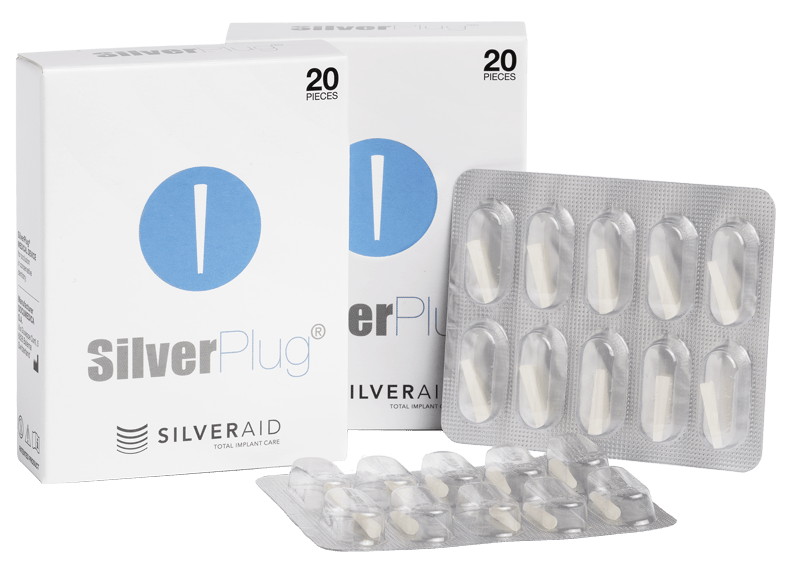
שבבי כסף SilverPlug

הוא המוצר הדגל של חברת Silveraid, שנועד לעזור לאנשי מקצוע בתחום רפואת השיניים במאמץ היומיומי שלהם לשמר תוצאות השיקום ולמנוע פרי-אימפלנטיטיס. מדובר באתגר: ניתן למנוע ולרפא את התופעה, שנחשבה לתופעת לוואי נורמלית, אומנם מטרידה ובלתי נמנעת של השתלות שיניים. הסיבה והמטרה הסופית של פיתוח המוצר היא בריאות טובה למטופלים ושקט נפשי לרופא המשתיל.

מדובר במכשיר הרפואי היחיד בעל פטנט עולמי, אשר מוסמך לאיטם מערכת השתלים הדנטליים. תוספת זאוליט כסף עם אפקט האנטי-בקטריאלי הטבעי שלו, מסייע במניעת זיהומים, דלקות, פרי-אימפלנטיטיס ועוד.
סילברפלאג - שבב כסף שמגן על השתלים שלך

אופן השימוש בסילברפלאג

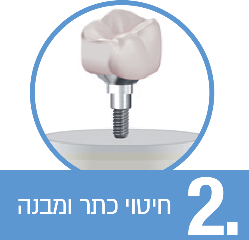
שבב הכסף סילברפלאג® נועד לאיטום פיר הכניסה לברגים של כתרים, גשרים ושיקומים מוברגים.

לאחר איטום באמצעות סילברפלאג, מומלץ להסתיר את פתח הגישה עם סיל-פלו, החומר מוקשה אור.
שבב הכסף - שימוש בשיקומים מוברגים

עלון מידע למטופל

כמה צעדים פשוטים בדרך אל בריאות השתל

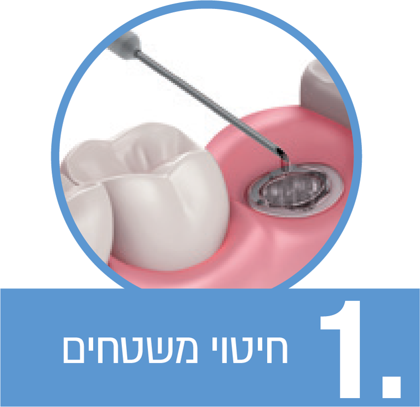
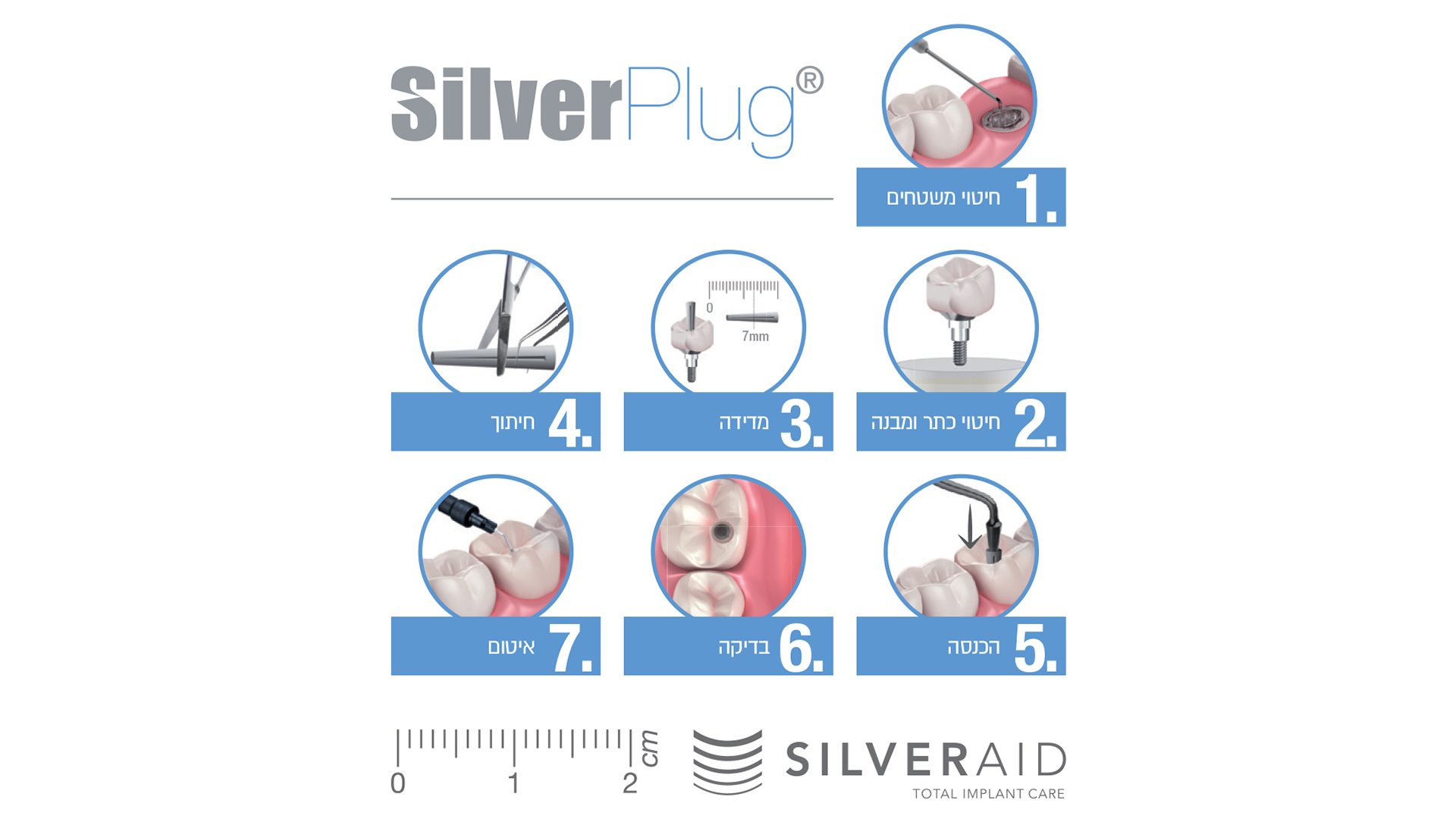
נקו את משטח השתל והסירו את הביופילם בעזרת מברשת גלילית.  יש לחטא היטב את האזור כולו באמצעות תמיסת חיטוי מתאימה. לאחר מכן, שטפו את האזור עם תמיסת מלח סטרילית.

כעת, נקו, הברישו וטבלו את המבנה בתמיסת חיטוי מתאימה למשך מספר דקות.

לאחר מכן, שטפו אותו היטב את בתמיסת סליין סטרילית.
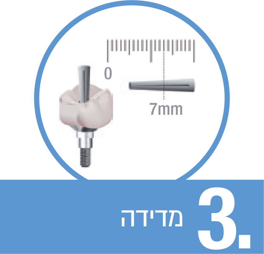
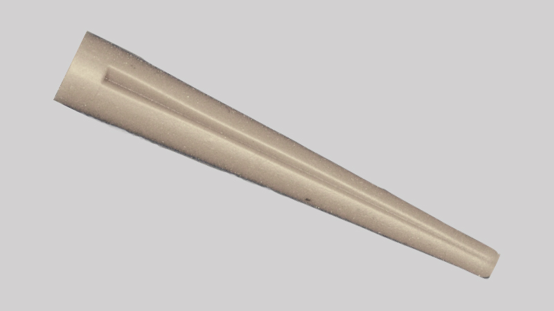
צורתו של סילברפלאג היא טרונקו-קוניקית, על מנת לאפשר את ההתאמה הנכונה של קוטרי נתיב הגישה למבנים.

כדי לעצב את סילברפלאג® לממדים הנכונים, וודאו את עומק הפיר הברגה במבנה, באמצעות מד עומק , והעריכו את הקוטר המרבי הנדרש. 
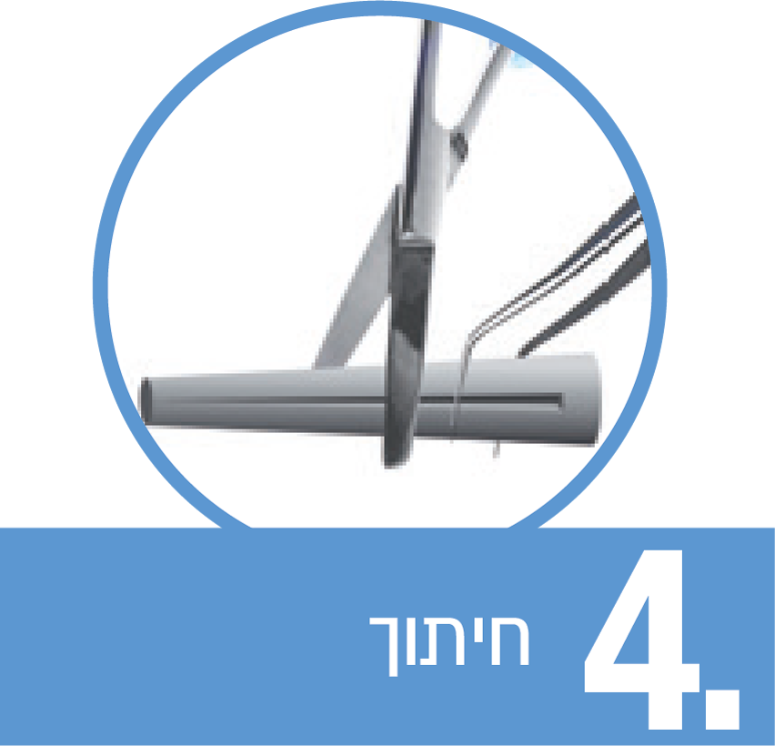
חתכו את סילברפלאג כך שימלא את נתיב הגישה ויהיה דבוק לחלוטין לדופן הצדדית ביותר שלו, בחלקו הגדול יותר.
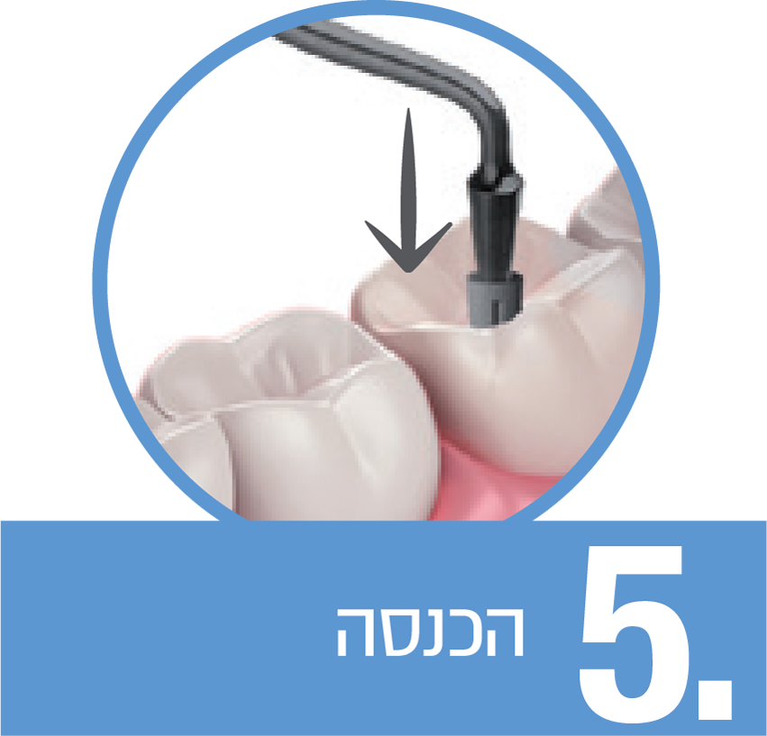
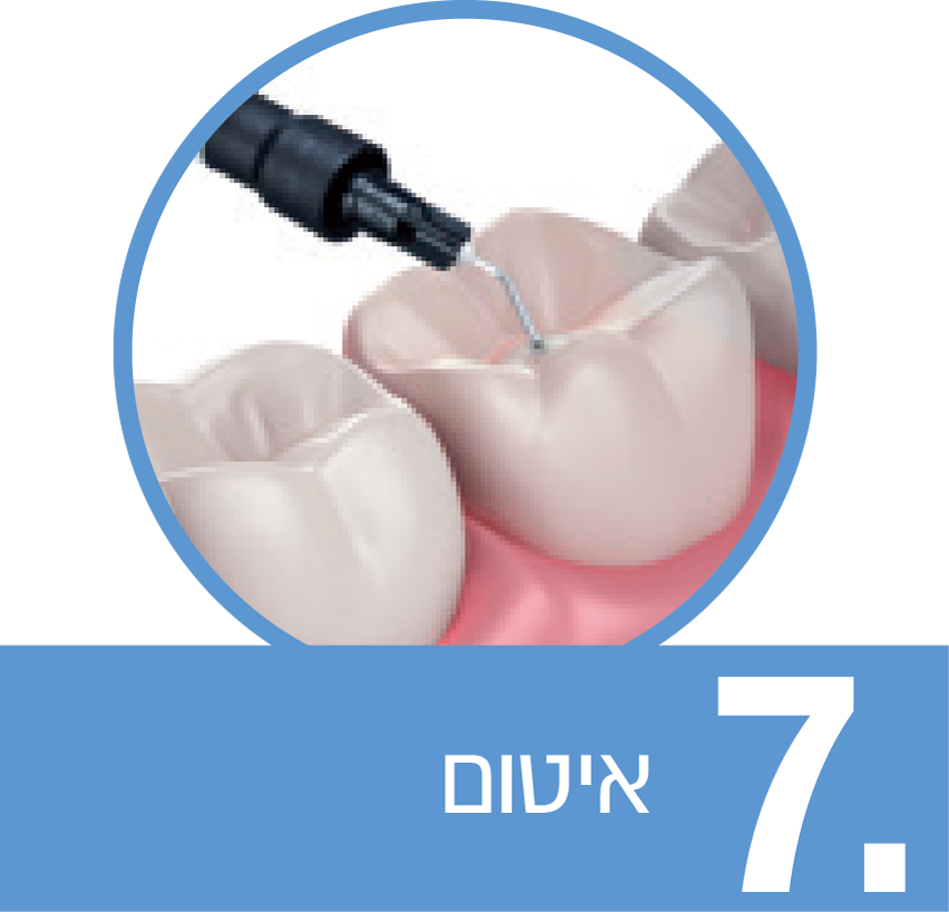
התקינו את השיקום על השתל והבריגו את הבורג לפי טורק הרצוי.  הצמידו את הסילברפלאג המוכן עם זונדה היעודית, והכניסו אותו בקלות בעזרת הפלאגר, הנמצא על הצד השני של הזונדה

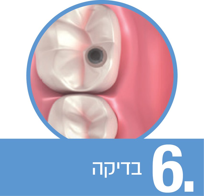
וודאו כי סילברפלאג אוטם לחלוטין את נתיב הגישה.

אם הסילברפלאג שהכנסתם ארוך מדי, אפשרי לקצץ אותו בעזרת מקדח יהלום

במידה ומדובר בכתר מוברג, זכרו להשאיר מקום לשכבה דקה של חומר איטום סילפלו..
לסיום, אטמו את פתח הכניסה עם חומר ייעודי מוקשה אור ("סילפלו"). ההמשך לפי פרוטוקול הסטנדרטי.

נקו את משטח השתל והסירו את הביופילם בעזרת מברשת גלילית.

 יש לחטא היטב את האזור כולו באמצעות תמיסת חיטוי מתאימה. לאחר מכן, שטפו את האזור עם תמיסת מלח סטרילית.

כעת, נקו, הברישו וטבלו את המבנה בתמיסת חיטוי מתאימה למשך מספר דקות.

לאחר מכן, שטפו אותו היטב את בתמיסת סליין סטרילית.

צורתו של סילברפלאג היא טרונקו-קוניקית, על מנת לאפשר את ההתאמה הנכונה של קוטרי נתיב הגישה למבנים.

כדי לעצב את סילברפלאג® לממדים הנכונים, וודאו את עומק הפיר הברגה במבנה, באמצעות מד עומק , והעריכו את הקוטר המרבי הנדרש. 

חתכו את סילברפלאג כך שימלא את נתיב הגישה ויהיה דבוק לחלוטין לדופן הצדדית ביותר שלו, בחלקו הגדול יותר.

התקינו את השיקום על השתל והבריגו את הבורג לפי טורק הרצוי.

 הצמידו את הסילברפלאג המוכן עם זונדה היעודית, והכניסו אותו בקלות בעזרת הפלאגר, הנמצא על הצד השני של הזונדה

וודאו כי סילברפלאג אוטם לחלוטין את נתיב הגישה.

אם הסילברפלאג שהכנסתם ארוך מדי, אפשרי לקצץ אותו בעזרת מקדח יהלום

  1. במידה ומדובר בכתר מוברג, זכרו להשאיר מקום לשכבה דקה של חומר איטום סילפלו..
לסיום, אטמו את פתח הכניסה עם חומר ייעודי מוקשה אור ("סילפלו"). ההמשך לפי פרוטוקול הסטנדרטי.

מדריכים שימושיים

סרטוני הדרכה פשוטים שיסייעו לכם לשלב את סילברפלאג בעבודה היומיומית שלכם

מאמרים קליניים

הנושא: חריצים, מיקרו-פערים, דליפות במערכות שתלים

Cone-morse connection implant system has very low bacterial leakage percentage and is similar to one-piece implants. למד עוד
Systematic review studies showed that there was some amount of microleakage at abutment implant interface. למד עוד
The present study confirms previous results about the high prevalence of bacterial penetration of screw-retained implant-abutment assemblies. למד עוד
The results of this study showed that, over time, bacteria will leak through the implant-abutment microgap at the implant-abutment interface. Implants with a titanium abutment demonstrate a smaller microgap than implants with a zirconia abutment. למד עוד
A newly developed test modelthat is a sensitive tool for the detection of differences between current implant systems with respect to their ability to prevent bacterial penetration at the implant-abutment interface under dynamic loading conditions. למד עוד
Systematic review studies showed that there was some amount of microleakage at abutment implant interface. למד עוד
The results of this study support the hypothesis that different types of implant-abutment joints are responsible for the observed differences in bacterial penetration. למד עוד
Gaps were sealed with the chlorhexidine-containing varnish Cervitec with a positive feedback. (in vistro study). למד עוד
Conclusion: While clone abutments may look similar to the original component, they display considerable differences and variations in their physico-mechanical characteristics detectable by advanced testing methods. How much these differences affect reliability and longevity of the restoration's clinical performance should be investigated in clinical studies. למד עוד
Premature loss of dental implants is due, apart from mechanical factors, to germrelated inflammation. Gaps and hollow spaces within the implant system, for example the gap between implant and abutment in the two-part implant system, may provide a bacterial reservoir causing or maintaining inflammation. The bacterial spectrum involved is similar to that found in periodontitis. This in vitro study aimed to scrutinise the capability of Porphyromonas gingivalis (DSM 20709), the bacterium blamed for inducing peri-implantitis, to pass the implant/abutment gap in titanium implant systems used for orthodontic anchorage and to remain vital in the interior. Additionally, the in vitro effectiveness of gutta percha for gap sealing was examined. Twelve titanium implants (Straumann, diameter: 3.3 mm, length 5.5 mm) were provided with abutments at a defined torque (20 Ncm), six of which were sealed with gutta percha before screwing in the abutment. Subsequently the implants were placed in a nutrient solution (thioglycolate boullion with haemin-menadione solution) that contained Porphyromonas gingivalis. Microbiological specimens were sampled from the implant interiors after 24 and 72 hours and analysed using culture methods. There was evidence that penetration of the periodontal pathogen Porphyromonas gingivalis to the implant interior may occur as early as after 24 hours. Microbes were also detected in the interior of implants sealed with gutta percha. The abutment/implant interface in vitro provides a microbiological leakage for the prospective peri-implantitis-inducing bacterium Porphyromonas gingivalis. Survival of the bacterium is possible in the interior, so that development of a bacterial reservoir is assumed. This in vitro trial produced no evidence that sealing with gutta percha is an effective means to prevent secondary bacterial colonisation in the implant interior. למד עוד
Conclusions: The reported results are better to those of previous studies carried out on different implant systems. Until now, none implant-abutment system has been proven to seal the gap between implant and abutment. למד עוד
Conclusion: The reported results are similar to previous work. Konus Implant System® showed bacterial leakage similar respect others implant systems (18% Porphiromonas Gingivalis, 19% Tannerella Forsitya versus 20% of Bicon© and Ankylos ® systems). In spite of the limits of our study, none two-piece implant system has been demonstrated to perfectly close the gap between implant and abutment. למד עוד
Different implant-abutment connections have been developed in the effort of reducing mechanical and biological failure. The most frequent complications are screw loosening, abutment or implant fracture and marginal bone loss due to overload and bacterial micro-leakage. Ideal connection should work as a one-piece implant avoiding the formation of a micro-gap at the implant-abutment interface. Different in vitro and in vivo researches have been published to compare the implant-abutment connections actually available: external hexagon, internal hexagon and conical finding different amount of micro-gap, micro-leakage and marginal bone loss. The aim of this article is to describe, according to the most recent literature, different kind of fixture-abutment connections and their clinical and mechanical advantages or disadvantages. למד עוד
Conclusion: On the basis of the results obtained in the present study using 2 different implant systems, we conclude that CRA implants offer better results relating to fluid and bacterial permeability compared to SRA implants. למד עוד
Conclusions: Ditron Implant IAC (MPI, Ditron Dental, Israel) is efficacy in reducing bacterial leakage. למד עוד
Conclusions: In vitro, S aureus leakage through the implant/abutment interface occurred with both Morse taper and internal-hexagon implants. למד עוד
Conclusion: FEA proved to be an adequate method for studying microgap formation in dental implant-abutment complexes. FEA is not limited in gap width resolution as are radiologic techniques and can also provide insight into stress distributions within the loaded complex. למד עוד
Conclusions: The abutment removal force or torque was not affected by mechanical cycling. Bacterial sealing of the implant/abutment tapered interface was not effective for any condition analyzed. Imprecise machining of implant parts does not allow a sufficient contact area between surfaces to provide effective sealing and prevent bacterial leakage. למד עוד
Conclusions: Implant's internal contamination shows that IAC is not sealing. The reported results are similar to those of previous studies carried out on different implant systems. Until now, no IAC has been proven to seal the gap between implant and abutment. למד עוד
Abstract Gaps and hollow spaces at the implant abutment interface will act as a bacterial reservoir that may cause peri-implantitis. Hence, the sealing ability of O-ring (in addition to polysiloxane) and GapSeal (an antibacterial sealing gel) was evaluated. A total of 45 identical implant systems (ADIN Dental Implant Systems) were divided into 3 groups of 15 implants each: an unsealed group, a group sealed with O-rings, and a group sealed with GapSeal gel. The implant and abutment were gamma sterilized after assembly. Two implants from each group were randomly incubated in sterile brain heart infusion (BHI) broth tubes and checked for sterility. The remaining 13 implants were incubated in BHI broth inoculated with Enterococcus and incubated for 5 days. They were then removed from the tubes, dried aseptically, placed in 2% sodium hypochlorite solution for 30 minutes, and washed with sterile saline for 5 minutes. Next, the assembly was dried aseptically and put in sterile BHI broth tubes and incubated for 24 hours to check surface sterility. Keeping 2 implants as controls from each group, the remaining 11 implants were dismantled group-wise and placed in liquid BHI broth; the test tubes were then shaken thoroughly so that the broth would come in contact with all implant surfaces. The solution from this tube was poured on pre-prepared sterile agar plates and incubated for 24 hours. The colonies formed on the agar plate were then counted using a digital colony counter. The data thus obtained were subjected to statistical analysis by Kruskal-Wallis analysis of variance and Mann-Whitney U test. It was concluded that though microbial growth is seen in all the 3 groups, the least growth was seen in the GapSeal group followed by the O-ring group. למד עוד
Conclusion: The determination of the volumes and the evaluation of leaked substance using stereomicroscopic and spectrophotometric methods showed the accuracy of these methods and the importance of their use in the study of microleakage. למד עוד
Conclusions: The data presented here demonstrate that dental implants fabricated with gap-free abutments using a shape memory alloy showed significantly reduced bacterial leakage versus conventional implants. This improvement could minimize clinical problems such as peri-implantitis and consequently enhance the long-term success of dental implants. למד עוד
Compare the leakage rates of different types of implant-abutment connections. למד עוד
Conclusion: There was a significant difference related to the strength of tightening. Under the conditions of our experiment, the gap of connection was reduced with a torque of ≥20 N cm. Only a small portion of the clinicians could obtain these values by hand. Therefore, a dynamometrical manual wrench should be used to minimize the gap during the osseointegration period. למד עוד
Compare the leakage rates of different types of implant-abutment connections. למד עוד

הנושא: רובד בקרטיאלי ודליפות בפלטפורמות שונות

Cone-morse connection implant system has very low bacterial leakage percentage and is similar to one-piece implants. למד עוד
Systematic review studies showed that there was some amount of microleakage at abutment implant interface. למד עוד
The present study confirms previous results about the high prevalence of bacterial penetration of screw-retained implant-abutment assemblies. למד עוד

הנושא: שימוש בחומרים שונים וכסף לאיטום חריצים

Conclusions: The casting retention on implant-abutments provided by COP was similar to that of copings cemented with zinc phosphate and may be influenced by the addition of calcium carbonate. למד עוד
Conclusions: No significant differences were found between alloy/index combinations, except for the measured variable dtheta(y), angular change about the Y axis. למד עוד
This report presents the use of a dental implant with an anti-rotational attachment for the retention of a thumb prosthesis. A retention system was manufactured with an attachment (UCLA) screwed into the implant with a two-bar system that was cast in metallic silver palladium. A substructure made from heat-cured acrylic resin was joined to the retention system by clips to join the thumb to the finger (bar clip) in the cast with implant rejoinder. The silicone material, Silastic-MDX 44210, was used to achieve function and aesthetics. Following osseointegration, no skin problems were observed. Whilst the implant-retained digital prosthesis presented some motor limitations, it allowed the patient to return to entertainment and achieve social conviviality. למד עוד
This paper discusses dental restorative resins, alloys, cements and implants and emphasizes the need for useful laboratory information that will predict the clinical behavior of these materials and structures. למד עוד
Study on rat tissue reaction to operatively inserted implants of different acrylic resin materials used in prosthetic dentistry. למד עוד
The aim of this study was to investigate the influence of different pins usually used for intraoperative root filling on periapical bone healing. In a specially designed animal experiment it was possible to evaluate biocompatibility simultaneously with the influence of varying materials on bone regeneration in standardized defects. Bioincompatible silver, acrylate and gutta percha root filling delay and impede bone regeneration. Biocompatible aluminium oxide ceramic and titanium pins do not have any influences on spontaneous bone regeneration. Reducing the defect, however, they do improve the conditions for complete bony regeneration. למד עוד

חוות דעת מומחים

תמיד עומדים לרשותכם ומוכנים לענות על כל שאלה שיש לכם בנוגע למוצרים